Leg Bone Diagram / Lower leg - bones | Diagram | Patient / Ct, mri, radiographs, anatomic diagrams and nuclear images.. The foot bones shown in this diagram are the talus, navicular, cuneiform, cuboid, metatarsals. The human leg, in the general word sense, is the entire lower limb of the human body, including the foot, thigh and even the hip or gluteal region. High resolution textures and displacement included. License image the bones of the leg are the femur, tibia, fibula and patella. 25.09.2018 · leg bone anatomy diagram diagram.
Foot bones illustration with icons. Want to learn more about it? Click now to learn more about the bones leg and knee anatomy: 25.09.2018 · leg bone anatomy diagram diagram. Your leg bones are the longest and strongest bones in your body.
Bones of the Lower Limb | Anatomy and Physiology from s3-us-west-2.amazonaws.com Bones give your body structure and enable you to move, but what else is your skeletal system responsible for? Download leg bone stock vectors. Joints of hand anterior view, lateral view, right hand. Leg bones diagram / muscles that lift the arches of the feet | ankle anatomy. Master leg and knee anatomy using our topic page. Most of the animals have the same bones, although some are shaped differently and placed in different positions. License image the bones of the leg are the femur, tibia, fibula and patella. Most relevant best selling latest uploads.
Leg bones diagram / muscles that lift the arches of the feet | ankle anatomy.
25.09.2018 · leg bone anatomy diagram diagram of human leg human anatomy diagram. Your leg bones are the longest and strongest bones in your body. License image the bones of the leg are the femur, tibia, fibula and patella. How to draw hand bone diagram| phalange carpal bone diagram. Posted on april 18, 2019april 18, 2019. Skeleton leg ankle joints and toe. Each leg is made up of four bones. Most relevant best selling latest uploads. The bones of the leg are the femur, tibia, fibula and patella. Affordable and search from millions of royalty free images, photos and vectors. 25.09.2018 · leg bone anatomy diagram diagram. Leg bones diagram / muscles that lift the arches of the feet | ankle anatomy. Horses engage in a lot of physical activity, and the consequences.
What does this suggest about mammals? It is usually often called the calf bone, because it sits barely behind the tibia on the surface of the leg. Click now to learn more about the bones leg and knee anatomy: 25.09.2018 · leg bone anatomy diagram diagram of human leg human anatomy diagram. Affordable and search from millions of royalty free images, photos and vectors.
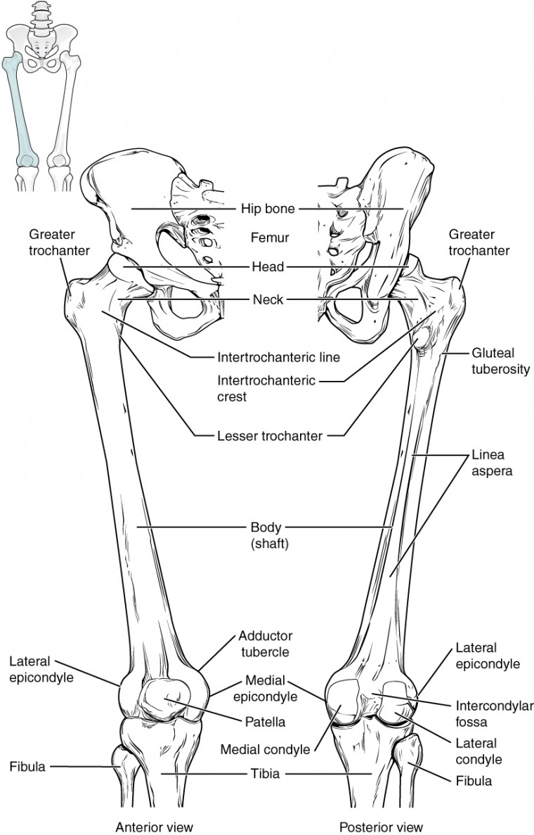
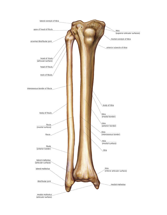
Diagram Of The Leg Bones Diagram Of Leg Bones Human Leg ... from i.pinimg.com In this image, you will find femur, medial epicondyle of the femur, patella, tibial tuberosity, anterior rest of. Master leg and knee anatomy using our topic page. The foot bones shown in this diagram are the talus, navicular, cuneiform, cuboid, metatarsals and calcaneus. This diagram with labels depicts and explains the details of bones of lower leg. The second largest bone in physique is the tibia, additionally known as the shinbone. Distal end of right humerus. Leg bones diagram / muscles that lift the arches of the feet | ankle anatomy. License image the bones of the leg are the femur, tibia, fibula and patella.
Most of the animals have the same bones, although some are shaped differently and placed in different positions.
Ct, mri, radiographs, anatomic diagrams and nuclear images. Bone circulatory disturbances in the development of spontaneous bacterial chondronecrosis with osteomyelitis: Anchor chart diagram leg human knee skeleton health bone science human body. How to draw hand bone diagram| phalange carpal bone diagram. Includes leg (femur, tibia, patella, and fibula) and foot (tarsals and digits) bones. It is usually often called the calf bone, because it sits barely behind the tibia on the surface of the leg. Learn vocabulary, terms and more with flashcards, games and other study tools. Human foot bones anatomy sketch of orthopedics medicine. Click now to learn more about the bones leg and knee anatomy: A translational model for the pathogenesis of femoral head necrosis. How to draw a human bone leg. The foot bones shown in this diagram are the talus, navicular, cuneiform, cuboid, metatarsals. Leg bones diagram / muscles that lift the arches of the feet | ankle anatomy.
Click now to learn more about the bones leg and knee anatomy: Most relevant best selling latest uploads. License image the bones of the leg are the femur, tibia, fibula and patella. High resolution textures and displacement included. Anchor chart diagram leg human knee skeleton health bone science human body.
Leg Bone Diagram : Picture Of Human Leg Bone Page 1 Line ... from images.fineartamerica.com What does this suggest about mammals? The radius and ulna are two parallel bones which extend from in four legged mammals ,the ulna is responsible for muscle attachment. Start studying leg bone labeling. Basic bone diagram wiring diagrams click, diagram of nephron simple horse muscle and bone skeleton leg image, simple bone diagram wiring diagram library, free printable dinosaur skeleton. Knee bone diagram illustrations & vectors. Your leg bones are the longest and strongest bones in your body. Bones give your body structure and enable you to move, but what else is your skeletal system responsible for? Foot bones illustration with icons.
Leg bone stock vectors, clipart and illustrations.
Human foot bones anatomy sketch of orthopedics medicine. High resolution textures and displacement included. Foot bones illustration with icons. What does this suggest about mammals? The foot bones shown in this diagram are the talus, navicular, cuneiform, cuboid, metatarsals. Learn vocabulary, terms and more with flashcards, games and other study tools. This diagram with labels depicts and explains the details of bones of lower leg. Skeleton leg ankle joints and toe. The foot bones shown in this diagram are the talus, navicular, cuneiform, cuboid, metatarsals. Horses engage in a lot of physical activity, and the consequences. Basic bone diagram wiring diagrams click, diagram of nephron simple horse muscle and bone skeleton leg image, simple bone diagram wiring diagram library, free printable dinosaur skeleton. A translational model for the pathogenesis of femoral head necrosis. How to draw a human bone leg.


0 Komentar